

For decades, scientists viewed the genome of a newly fertilized egg as a structural “blank slate” – a disordered tangle of DNA waiting for the embryo to “wake up” and start reading its own genetic instructions. In research published February 24, 2026 in Nature Genetics, Professor Juanma Vaquerizas and his team have found that a surprising level of structure is already in place. They’ve developed a breakthrough technology, called Pico-C, which enables scientists to see the 3D structure of the genome in unprecedented detail. Using this technique, they discovered that well before the genome fully awakens – a critical event known as Zygotic Genome Activation – a sophisticated 3D scaffold of DNA is already being built. Understanding how DNA folds in space matters because this controls which genes can be turned on during development, helping cells function properly and preventing developmental defects and disease. The open-access article is titled “Three-Dimensional Genome Reorganization Foreshadows Zygotic Genome Activation in Drosophila.” “We used to think of the time before the genome awakens as a period of chaos,” explains Noura Maziak, lead author of the study. “But by zooming in closer than ever before, we can see that it’s actually a highly disciplined construction site. The scaffolding of the genome is being erected in a precise, modular way, long before the ‘on’ switch is fully flipped.”
Pico-C: Seeing More with Less
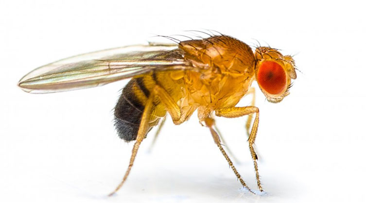
The team’s discovery was made in the fruit fly (Drosophila). In the first few hours after fertilization, a fly embryo undergoes a rapid series of nuclear divisions, creating thousands of cells. It’s this high-speed environment that makes the fruit fly perfect for studying the fundamentals of genetics.
Using their ultra-sensitive Pico-C technology, the researchers mapped the 3D structure of the fruit fly genome during these early developmental stages. They found that the 3D loops and folds of DNA follow a modular logic, allowing different inputs to regulate specific parts of the genome. It is a complex architectural program that ensures the information encoded in the genome is ready for action the moment it is needed.
As well as providing high-resolution detail about the shape of DNA, Pico-C only requires tiny amounts of sample – ten times less than standard methods. This opens the door to opportunities for studying how DNA folding shapes gene regulation and its implication in many diseases in greater detail than previously possible.
From Fly Embryos to Human Health
While the “blueprint” of this architecture was discovered in fruit flies, the importance of maintaining it applies directly to humans. In a companion study published February 24, 2026 in Nature Cell Biology, led by Professor Ulrike Kutay and collaborators at ETH Zürich in Switzerland, the team applied this high-resolution mapping to human cells. That open-access article is titled “LBR and LAP2 Mediate Heterochromatin Tethering to the Nuclear Periphery to Preserve Genome Homeostasis.”
They investigated what happens when the “anchors” that hold this 3D structure in place are removed. The results were striking: when the architecture collapses, the human cell mistakes the structural failure for a viral attack. This triggers the cell’s innate immune system, sounding a false alarm that can lead to inflammation and disease.
“These two studies tell a complete story,” says Vaquerizas. “The first shows us how the genome’s 3D structure is carefully built at the start of life. The second shows us the disastrous consequences for human health if that structure is allowed to collapse.”
[News release] [Nature Genetics article] [Nature Cell Biology article]