

Researchers uncover a central molecular pathway linking vesicle trafficking to cancer immune escape, offering a promising new therapeutic target.
by Manisha Kashyap, PhD
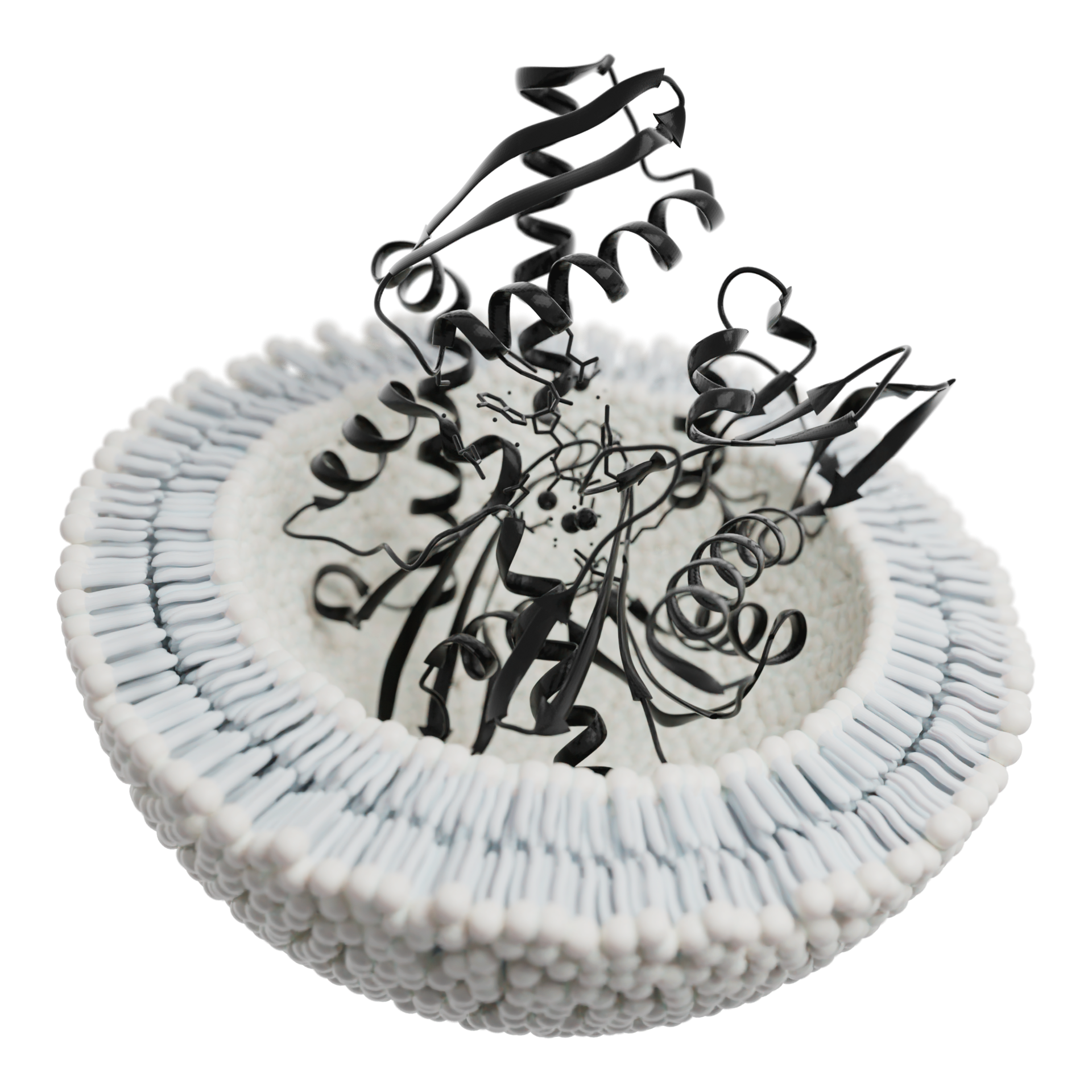
A collaborative team from Huazhong University of Science and Technology and the Guangzhou Institutes of Biomedicine and Health in China has revealed how tumors exploit exosomal transport machinery to suppress the immune system. Their open-access study, published in Nature Communications (October 13, 2025), under the title “Munc13-4 Mediates Tumor Immune Evasion by Regulating the Sorting and Secretion of PD-L1 Via Exosomes;” identifies Munc13-4 as a pivotal regulator of programmed death-ligand 1 (PD-L1) sorting and secretion through exosomes, uncovering a powerful mechanism of tumor immune evasion and suggesting a new avenue to enhance the effectiveness of immunotherapy.
Exosomal PD-L1 and Resistance to Immunotherapy
Tumor cells evade immune destruction primarily by expressing PD-L1, which binds to PD-1 receptors on T cells and suppresses their cytotoxic activity. While PD-1/PD-L1 checkpoint inhibitors have shown remarkable clinical success, many patients remain unresponsive. A major reason, the authors note, is that tumors also release PD-L1-containing exosomes—tiny extracellular vesicles (EVs) that circulate systemically, inhibiting T cell function far from the tumor site. Understanding how PD-L1 is packaged into these vesicles has been a critical gap in cancer immunology.
Munc13-4: A Crucial Vesicle-Trafficking Regulator
Through genomic and proteomic analysis, the researchers found that Munc13-4—a member of the Munc13 protein family involved in vesicle exocytosis—is upregulated across multiple cancer types, including breast, thyroid, pancreatic, and liver cancers.
Using CRISPR-Cas9 gene editing, the scientists deleted Munc13-4 in murine and human breast cancer cell lines. The knockout did not alter cell proliferation in vitro, but in animal models it caused a dramatic reduction in tumor growth.
This effect depended on an intact immune system: Munc13-4-deficient tumors grew slowly in immunocompetent mice but regained aggressiveness in T-cell-deficient hosts, confirming that Munc13-4 promotes tumor progression by suppressing immune responses.
Enhanced T-Cell Infiltration and Activation
Loss of Munc13-4 led to a striking increase in CD4⁺ and CD8⁺ T-cell infiltration in tumors, spleens, and lymph nodes. These T cells showed higher expression of activation markers such as granzyme B, Ki67, and interferon-γ (IFNγ), indicating stronger proliferation and cytotoxicity. The findings demonstrate that Munc13-4 deficiency reinvigorates systemic T-cell-mediated anti-tumor immunity.
Mechanistic Basis: PD-L1 Secretion Via Exosomes
Although total PD-L1 and cell-surface PD-L1 levels were unchanged, exosomal PD-L1 secretion dropped sharply after Munc13-4 knockout. Electron microscopy and nanoparticle tracking confirmed reduced exosome release.
When exosomes from wild-type tumor cells were transferred into mice bearing Munc13-4-deficient tumors, tumor growth accelerated, but this effect was abolished when PD-L1 was neutralized.
Thus, Munc13-4 controls PD-L1 export through exosomes, directly influencing tumor-induced immune suppression.
Synergy with Immune Checkpoint Blockade
Tumors lacking Munc13-4 were markedly more responsive to anti-PD-1 and anti-PD-L1 antibody therapy than controls. In Munc13-4-competent tumors, checkpoint blockade produced little benefit, but combining it with Munc13-4 loss significantly delayed tumor growth, suggesting that targeting Munc13-4 could overcome immunotherapy resistance.
Molecular Interactions: Rab27, SNAREs, and HRS
At the mechanistic level, Munc13-4 collaborates with the small GTPase Rab27a and SNARE proteins (syntaxin-4, SNAP-23, VAMP-7) to enable docking and fusion of multivesicular bodies (MVBs) with the plasma membrane. Cryo-electron microscopy at 3.4 Å resolution revealed structural details of the Munc13-4–Rab27a complex, clarifying how it stabilizes vesicle docking before exosome release. Munc13-4 also interacts with HRS, a key ESCRT-0 component responsible for endosomal cargo sorting. Together, Munc13-4, HRS, and PD-L1 form a ternary complex that ensures PD-L1 is directed into exosomes rather than degraded in lysosomes.
IFNγ-Dependent Modifications Fine-Tune PD-L1 Sorting
Interferon-γ (IFNγ) signaling—an important mediator of anti-tumor immunity—was shown to modulate Munc13-4 and HRS through opposing post-translational changes:
–Munc13-4 acetylation, catalyzed by the CBP acetyltransferase, weakened its binding to PD-L1 and inhibited PD-L1 sorting.
–HRS deubiquitylation, regulated by enzymes such as NEDD4L and USP11, strengthened HRS–PD-L1 interaction and enhanced PD-L1 packaging into exosomes.
This dynamic regulation allows tumor cells to exploit immune signaling pathways to balance PD-L1 production and secretion.
Therapeutic Strategy: Blocking the Munc13-4–PD-L1 Interaction
To test whether interrupting this pathway could restore immune surveillance, the authors developed a synthetic peptide that specifically blocks the Munc13-4–PD-L1 interaction. Treatment with this peptide impeded PD-L1 sorting, enhanced T-cell cytotoxicity, and suppressed tumor growth in vivo. The results highlight Munc13-4 as a druggable target whose inhibition could synergize with existing immune checkpoint therapies.
Discussion
Tumor-derived exosomes carrying PD-L1 play a pivotal role in immune evasion by binding to PD-1 receptors on T cells, suppressing immune activity both within the tumor and at distant sites. This study identifies Munc13-4 as a critical regulator of this process, demonstrating that its deletion reduces exosomal PD-L1 secretion, enhances T cell-mediated anti-tumor responses, and improves the effectiveness of immune checkpoint inhibitors. Mechanistically, Munc13-4 forms complexes withHRS, Rab27a, and SNARE proteins to coordinate PD-L1 sorting, MVB docking, and exosome release. Moreover, IFNγ signaling modulates Munc13-4 and HRS through acetylation and deubiquitylation, respectively, fine-tuning PD-L1 packaging into exosomes. These findings establish Munc13-4 as a key mediator linking vesicle trafficking to tumor immune suppression and highlight its potential as a therapeutic target to enhance immunotherapy efficacy.
Conclusion
This landmark study identifies Munc13-4 as a master regulator connecting vesicle-trafficking machinery to immune checkpoint signaling. By facilitating PD-L1 export via exosomes, Munc13-4 enables tumors to escape immune control both locally and systemically. Disrupting this pathway boosts T-cell activity, sensitizes tumors to PD-1/PD-L1 blockade, and markedly limits tumor progression. Targeting Munc13-4—or its interaction with PD-L1—represents a promising strategy to overcome immune evasion and enhance the durability of cancer immunotherapy responses.
[Nature Communications article]
About the BioQuick Writer

Dr. Manisha Kashyap (Dr. MK)
BDS (Hons), MSc, PhD
Manisha Kashyap earned a BS in Dentistry (Gold Medalist) and an MS in Clinical Research, and she is a driven PhD scholar in Health Policy and Management.
With a rich background as a Drug Safety Analyst for top-tier bio pharmaceutical companies, she blends clinical expertise with a passion for advancing healthcare accessibility and innovation through her writing career for the love of science and technology via MedKnow Research Writing Solutions linkedin.com/in/medknowresearchwritingsolutionsbydrmk
Dr. Kashyap’s PhD thesis is on “Artificial Intelligence (AI) and Public Health: Harnessing the Power of AI for Enhanced Public Health Policy and Management.”